All published articles of this journal are available on ScienceDirect.

Development and Evaluation of a Topical Antibacterial Gel from Rhizophora apiculata Blume Leaves: Standardization, Phytochemical Quantification, and Bioactivity Assessment
 iD
David Tjahyadi2
iD
Hosea Jaya Edy3
iD
Joey Joshua Tjahyadi4
iD
Laurentia Gabrielle4
iD
Ashaolu Victoria Oladimeji5
iD
Seçil Karahüseyin6
iD
iD
David Tjahyadi2
iD
Hosea Jaya Edy3
iD
Joey Joshua Tjahyadi4
iD
Laurentia Gabrielle4
iD
Ashaolu Victoria Oladimeji5
iD
Seçil Karahüseyin6
iD
Authors Info & Affiliations
Abstract
Introduction
Rhizophora apiculata Blume is a mangrove species rich in polyphenolic secondary metabolites, including flavonoids, phenolic acids, and tannins, which are known to exhibit antibacterial activity. The development of topical gel formulations containing these bioactive compounds may provide a promising alternative approach for the management of skin and wound infections. This study aimed to develop and evaluate a standardized topical gel formulation containing R. apiculata Blume leaf extract and to assess its physicochemical characteristics, phytochemical content, and antibacterial activity.
Methods
Ethanolic leaf extract of R. apiculata Blume was incorporated into gel formulations at concentrations of 3%, 5%, and 7% (w/w). The resulting formulations were evaluated for physicochemical properties, including organoleptic characteristics, homogeneity, spreadability, adhesiveness, pH, and viscosity. Quantitative determination of flavonoids (Quercetin Equivalent Flavonoids, QEF), total phenolics (Gallic Acid Equivalent Phenolics, GAEP), and tannins (Tannic Acid Equivalent Tannins, TAET) was performed using UV–Vis spectrophotometry. Antibacterial activity was assessed against Staphylococcus aureus, Staphylococcus epidermidis, Escherichia coli, and Pseudomonas aeruginosa using the agar well diffusion method, with Bioplacenton® gel as a positive control. Data were analyzed using one-way analysis of variance (ANOVA) followed by Tukey’s HSD post hoc test, with p < 0.05 considered statistically significant.
Results
All formulations exhibited acceptable physicochemical characteristics, including homogeneous yellowish-green appearance, characteristic odor, spreadability ranging from 4.36–4.90 cm, adhesiveness of 31.67–38.50 s, skin-compatible pH values (6.36–6.70), and viscosity ranging from 125.83–175.00 Pa·s. Quantitative phytochemical analysis demonstrated a concentration-dependent increase in flavonoid, phenolic, and tannin contents, with the 7% formulation showing the highest QEF (0.5898 ± 0.0025 mg QE/g gel), GAEP (0.4395 ± 0.0067 mg GAE/g gel), and TAET (0.2845 ± 0.0025 mg TAE/g gel) values (p < 0.001). All formulations exhibited antibacterial activity against the tested bacterial strains. The 7% gel formulation produced the largest inhibition zones against E. coli (18.02 ± 0.41 mm), S. aureus (14.94 ± 0.30 mm), S. epidermidis (13.82 ± 0.27 mm), and P. aeruginosa (24.76 ± 0.21 mm), indicating enhanced antibacterial efficacy with increasing extract concentration.
Discussion
The observed concentration-dependent increase in antibacterial activity may be associated with the higher levels of flavonoids, phenolics, and tannins present in the formulations. The favorable physicochemical properties of the gels further support their suitability for topical application. Among the tested formulations, the 7% gel demonstrated the most promising overall performance in terms of phytochemical content and antibacterial activity.
Conclusion
R. apiculata Blume leaf extract was successfully formulated into a topical gel with desirable physicochemical characteristics and significant antibacterial activity against both Gram-positive and Gram-negative bacteria. The findings support the potential application of R. apiculata leaf extract as a natural source for topical antibacterial formulations, with the 7% formulation showing the most favorable characteristics and warranting further investigation.
1. INTRODUCTION
The Indonesian coastline, stretching across thousands of kilometers, supports extensive mangrove ecosystems of significant ecological value. These ecosystems are broadly recognized for their biodiversity, which provides various benefits to human populations. Strategic optimization of coastal biological resources can be achieved by exploring specific flora, such as Rhizophora apiculata Blume, a mangrove species valued for its secondary metabolites with traditional medicinal applications. Indigenous to the coastal regions of North Sulawesi, R. apiculata Blume has been the subject of recent phytochemical studies. Research indicates that leaf extracts derived from specimens collected in this area contain phenolic compounds, flavonoids, and tannins, suggesting their potential utility as components in traditional medicinal preparations [1]. In addition to their ecological roles, mangrove ecosystems harbor diverse bioactive compounds that remain underexplored for pharmaceutical applications. This potential is particularly relevant in tropical regions, where plant-based traditional medicine continues to play an essential role in community healthcare [2].
Among the diverse mangrove flora, R. apiculata Blume (family Rhizophoraceae) emerges as a species of particular interest due to its widespread distribution across Southeast Asian coastal ecosystems, including Indonesia. This species exhibits remarkable ecological adaptability and commonly thrives in intertidal mangrove environments characterized by high salinity and tidal fluctuations [3]. Intertidal mangrove habitats are characterized by extreme environmental conditions, including high salinity and regular tidal fluctuations. These stress factors induce oxidative stress in mangrove plants. In response, R. apiculata Blume develops adaptive biochemical defense mechanisms. These mechanisms promote the biosynthesis of protective secondary metabolites with potential therapeutic relevance [4].
Comprehensive phytochemical investigations of R. apiculata Blume have revealed a rich profile of bioactive constituents. Analysis of leaf and root extracts from specimens collected in the Pichavaram mangrove forest of Tamil Nadu, India, identified multiple compound classes including alkaloids, saponins, phenolics, tannins, flavonoids, and terpenoids [5]. These phytochemicals, particularly the polyphenolic compounds (flavonoids, phenolic acids, and condensed tannins), have demonstrated significant pharmacological properties including antibacterial, antioxidant, anti-inflammatory, and wound-healing activities [6]. Furthermore, Rhizophoraceae mangroves, including R. apiculata Blume, have been reported to exhibit significant antibacterial and therapeutic potential, which is associated with their diverse phytochemical composition [7].
The global resurgence of interest in plant-derived therapeutics, driven by concerns over antibiotic resistance, synthetic drug side effects, and sustainable healthcare solutions, has intensified research into natural product development [8]. Previous studies have demonstrated the successful development and evaluation of stable topical herbal formulations with antibacterial properties [9]. In addition, R. apiculata Blume leaf extracts have shown significant antimicrobial activity and bioactive potential, supporting their suitability for further pharmaceutical formulation development [10]. Building upon this foundation, we identified R. apiculata Blume leaf extract as a promising candidate for advanced topical formulation development, given its analogous phytochemical profile and documented bioactivity.
Topical semisolid dosage forms, particularly hydrogels, represent an optimal delivery system for plant extracts due to their favorable biopharmaceutical characteristics. Gels-defined as semisolid systems consisting of either suspended inorganic particles or hydrated three-dimensional polymeric networks within an aqueous or hydroalcoholic continuous phase-offer distinct advantages for cutaneous application [11]. These include enhanced patient compliance through ease of application, non-greasy aesthetic properties, favorable skin feel, and the ability to provide controlled release of incorporated actives while maintaining a moist wound environment conducive to healing [12]. Successful precedents exist for plant-based gels, such as formulations containing Azadirachta indica leaf extracts that have demonstrated efficacy in wound healing models at concentrations typically below 10% w/w [12, 13]. Hydroxypropyl methylcellulose (HPMC) was incorporated into the formulation as a viscosity-enhancing and film-forming polymer to improve gel consistency, physical stability, spreadability, and retention on the skin surface. In combination with Carbopol 940, HPMC contributes to the formation of a stable semisolid matrix suitable for topical applications.
Despite extensive documentation of the phytochemical composition and biological activities of R. apiculata Blume, most previous studies have primarily focused on crude extract characterization, antioxidant activity, antimicrobial screening, and toxicity evaluation [1, 5, 6, 10]. Comparatively fewer investigations have addressed the development of standardized topical formulations incorporating R. apiculata Blume extracts, particularly formulations supported by comprehensive physicochemical characterization, quantitative phytochemical standardization, and antibacterial performance evaluation. Furthermore, limited information is available regarding the relationship between phytochemical content and antibacterial efficacy in formulated semisolid dosage forms.
Therefore, the present study was designed to bridge the gap between phytochemical discovery and pharmaceutical product development by formulating and evaluating a standardized topical gel containing R. apiculata Blume leaf extract. The novelty of this study lies in the integration of formulation development, physicochemical characterization, quantitative determination of major antibacterial phytochemical groups (flavonoids, phenolics, and tannins), and antibacterial evaluation within a single standardized topical preparation. This integrated approach provides a more comprehensive assessment of the pharmaceutical potential of R. apiculata Blume leaf extract for topical applications.
The study was carried out through three integrated objectives. The first objective was the development and physicochemical characterization of a Carbopol-based hydrogel containing R. apiculata Blume leaf extract gel (Ra-leg). The extract was prepared at three concentrations, namely 3%, 5%, and 7% w/w. Polymer selection, pH adjustment, and preservation were optimized to ensure stability and biocompatibility. The second objective focused on formulation standardization. This included physicochemical evaluations such as organoleptic properties, homogeneity, spreadability, adhesiveness, pH, and rheological behavior. In addition, phytochemical analysis quantified key antibacterial compounds. Flavonoids were expressed as quercetin equivalent (QE), phenolics as gallic acid equivalent (GAE), and tannins as tannic acid equivalent (TAE). All analyses were performed using validated spectrophotometric methods. The third objective was an in vitro antibacterial evaluation. The activity of Ra-leg was tested against Gram-positive bacteria (Staphylococcus aureus and Staphylococcus epidermidis) and Gram-negative bacteria (Escherichia coli and Pseudomonas aeruginosa). The study aimed to determine concentration-dependent effects and establish dose–response relationships.
Overall, the findings are expected to provide scientific evidence supporting the development of standardized R. apiculata Blume leaf extract gel formulations and contribute to the growing body of knowledge on mangrove-derived pharmaceutical products. The results may also serve as a foundation for further studies involving advanced phytochemical characterization, stability assessment, and preclinical evaluation of plant-based topical antibacterial formulations.
2. MATERIALS AND METHODS
2.1. Research Design
This laboratory-based experimental study was designed to develop, standardize, and evaluate topical antibacterial gel formulations containing R. apiculata Blume leaf extract (Ra-leg). The study was conducted through sequential experimental phases consisting of: (i) collection and extraction of R. apiculata Blume leaves; (ii) preparation of gel formulations containing 3%, 5%, and 7% (w/w) leaf extract; (iii) physicochemical standardization of the gel formulations, including organoleptic evaluation, homogeneity, pH, spreadability, adhesiveness, and viscosity; (iv) quantitative determination of total flavonoid, total phenolic, and total tannin contents using UV–Visible spectrophotometry; (v) evaluation of antibacterial activity against representative Gram-positive and Gram-negative bacterial strains using the agar well diffusion method; and (vi) statistical analysis of all experimental data to determine significant differences among formulations.
The overall experimental workflow is presented in Fig. (1).
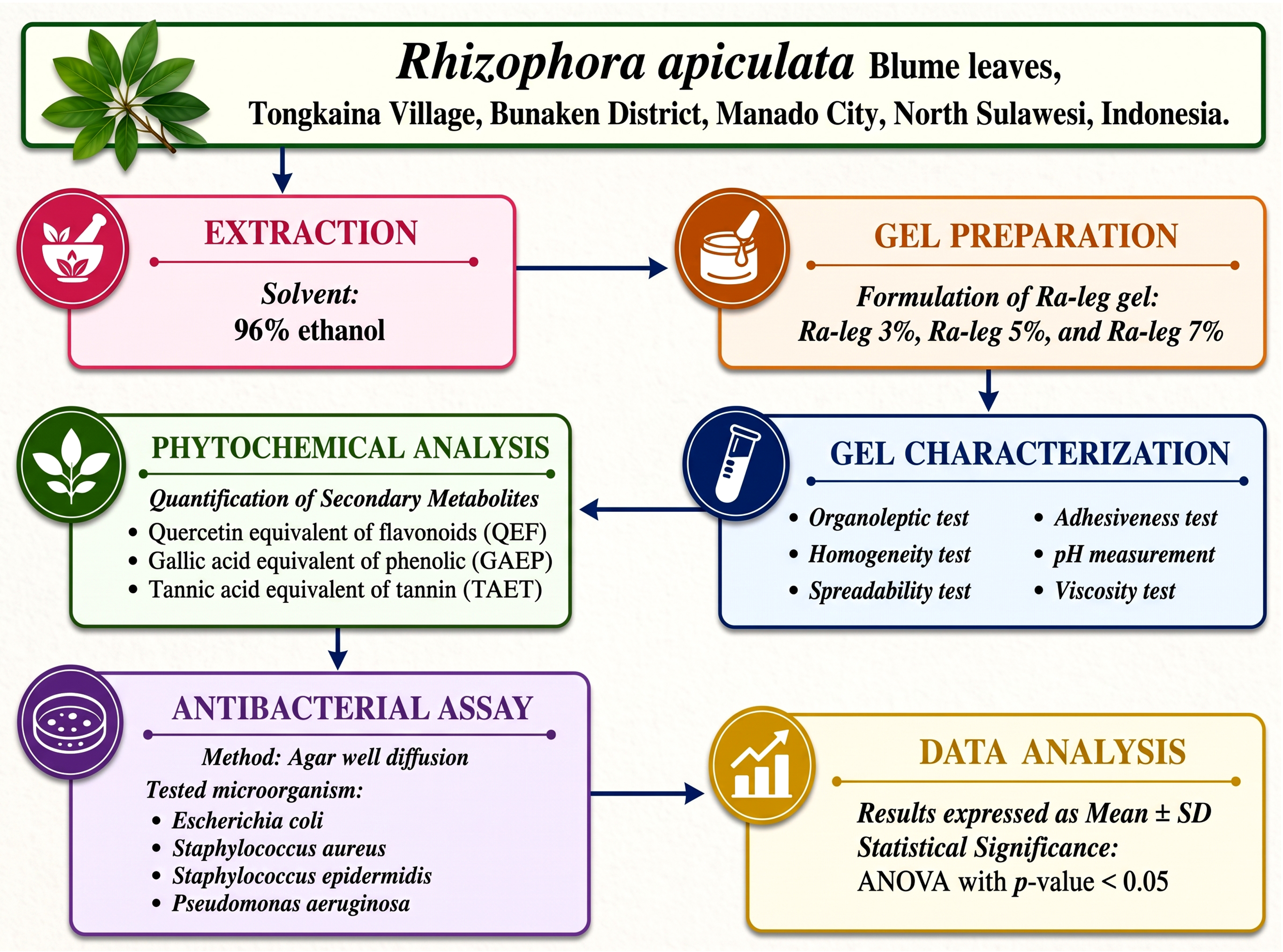
Research workflow of Ra-leg. Ra-leg: Rhizophora apiculata Blume leaf extract gel; SD: standard deviation.
2.2. Preparation of R. apiculata Blume Leaf Extract Gel
Leaves of Rhizophora apiculata Blume were collected from mangrove forests in Tongkaina Village, Bunaken District, Manado City, North Sulawesi, Indonesia. The collected leaves were washed thoroughly with clean water to remove adhering debris and dried under shaded sunlight using a black paranet until a constant weight was achieved. The dried leaves were ground into powder and extracted with 96% ethanol. The extract was concentrated using a rotary evaporator to obtain a viscous extract.
The concentrated extract was incorporated into topical gel formulations at concentrations of 3%, 5%, and 7% (w/w), designated as Ra-leg. Hydroxypropyl methylcellulose (HPMC) was used as the gelling agent to provide appropriate viscosity, spreadability, and physical stability of the formulation, thereby ensuring suitable consistency for topical application. The remaining excipients were incorporated according to the formulation composition, and the gel was mixed until a homogeneous preparation was obtained.
2.3. Gel Standardization
Standardization of Ra-leg was conducted by evaluating its physicochemical properties, including organoleptic characteristics, homogeneity, spreadability, adhesiveness, pH, and viscosity. Organoleptic properties (appearance, color, and odor) were assessed by direct observation. Homogeneity was evaluated by spreading the gel on a glass slide to ensure uniformity and the absence of coarse particles. The pH of the gel was measured using a calibrated pH meter by immersing the electrode into the gel until a stable reading was obtained.
Spreadability was determined by placing 0.5 g of gel between two glass plates. The spread diameter was measured after 1 min. A 150 g load was then applied for an additional 1 min. A spread diameter of 4–7 cm was considered indicative of acceptable semisolid consistency. Adhesiveness was measured by placing 500 mg of gel between two glass plates under a 200 g load for 2 min, and the time required for separation was recorded. Gel viscosity was measured using a Brookfield viscometer.
2.4. Determination of Secondary Metabolite Contents
Quantitative analysis of secondary metabolites in Ra-leg formulations (3%, 5%, and 7%) was performed using ultraviolet–visible (UV–Vis) spectrophotometry. Total flavonoid content was determined using quercetin as the reference standard and expressed as quercetin equivalent flavonoids (QEF). Total phenolic content was measured using gallic acid as the reference standard and expressed as gallic acid equivalent phenolics (GAEP). Total tannin content was determined using tannic acid as the reference standard and expressed as tannic acid equivalent tannins (TAET). Absorbance was measured at 510 nm for QEF, 650 nm for GAEP, and 740 nm for TAET.
2.5. Antibacterial Activity Assay
The antibacterial activity of Ra-leg was evaluated using the agar well diffusion method [13]. This method is widely used for screening the antimicrobial activity of plant extracts and topical formulations. The test organisms included E. coli, S. aureus, S. epidermidis, and P. aeruginosa. The bacterial strains used in this study were obtained from the American Type Culture Collection (ATCC). The following reference strains were used: Escherichia coli ATCC 25922, Staphylococcus aureus ATCC 25923, Staphylococcus epidermidis ATCC 12228, and Pseudomonas aeruginosa ATCC 27853. These bacteria represent both Gram-positive and Gram-negative pathogens commonly used in antibacterial susceptibility studies [14]. The assay was performed in five replicates. Plates were incubated at 37 °C for 24 hours before measuring inhibition zones using a digital caliper. Antibacterial activity was determined by measuring the diameter of the inhibition zones formed around the wells.
2.6. Data Analysis
Data obtained from gel standardization tests, secondary metabolite measurements, and antibacterial assays were expressed as mean ± standard deviation (SD). Data normality and homogeneity of variance were evaluated using the Shapiro–Wilk and Levene’s tests, respectively. Statistical differences among groups were analyzed using one-way analysis of variance (ANOVA), followed by Tukey’s HSD post hoc test for multiple comparisons. A p-value < 0.05 was considered statistically significant.
3. RESULTS
3.1. Ra-leg Composition
The composition of the Ra-leg formula is presented in Table 1.
| Components | Ra-leg | ||
|---|---|---|---|
| R. apiculata Blume leaf extract | 3% | 5% | 7% |
| Carbopol 940 | 2 | 2 | 2 |
| Hydroxypropyl methylcellulose (HPMC) | 2 | 2 | 2 |
| Polyethylene glycol 200 | 5 | 5 | 5 |
| Propylene glycol | 15 | 15 | 15 |
| Methyl paraben | 0.1 | 0.1 | 0.1 |
| Propylparaben sodium salt | 0.05 | 0.05 | 0.05 |
| Triethanolamine/sodium hydroxide 10% (q.s) | q.s. ad pH 6.5 | q.s. ad pH 6.5 | q.s. ad pH 6.5 |
| Aqua bidestillata | q.s. ad 100 g | q.s. ad 100 g | q.s. ad 100 g |
Abbreviations: Ra-leg: R. apiculata Blume leaf extract gel; q.s.: quantum satis (sufficient quantity).
3.2. Ra-leg Properties
Organoleptic and homogeneity tests of Ra-leg are presented in Table 2.
| Type of Organoleptic | Ra-leg | ||
|---|---|---|---|
| 3% | 5% | 7% | |
| Shape | Semi solid | Semi solid | Semi solid |
| Color | Transparent yellowish green | Transparent yellowish green | Transparent yellowish green |
| Odor | Characteristics of R. apiculata Blume leaf extract | Characteristics of R. apiculata Blume leaf extract | Characteristics of R. apiculata Blume leaf extract |
| Homogeneity | Homogeneous | Homogeneous | Homogeneous |
Abbreviations: Ra-leg: Rhizophora apiculata Blume leaf extract gel; cm: centimeter; SD: standard deviation; Pa·s: pascal second.
Table 2 presents the organoleptic and homogeneity characteristics of Ra-leg formulations. All formulations exhibited a semi-solid physical form with a transparent yellowish-green appearance and a characteristic odor of R. apiculata Blume leaf extract. All formulations were also homogeneous, indicating that the extract was uniformly dispersed throughout the gel base.
The results of the spreadability, adhesion, pH, and viscosity tests of Ra-leg are presented in Fig. (2).
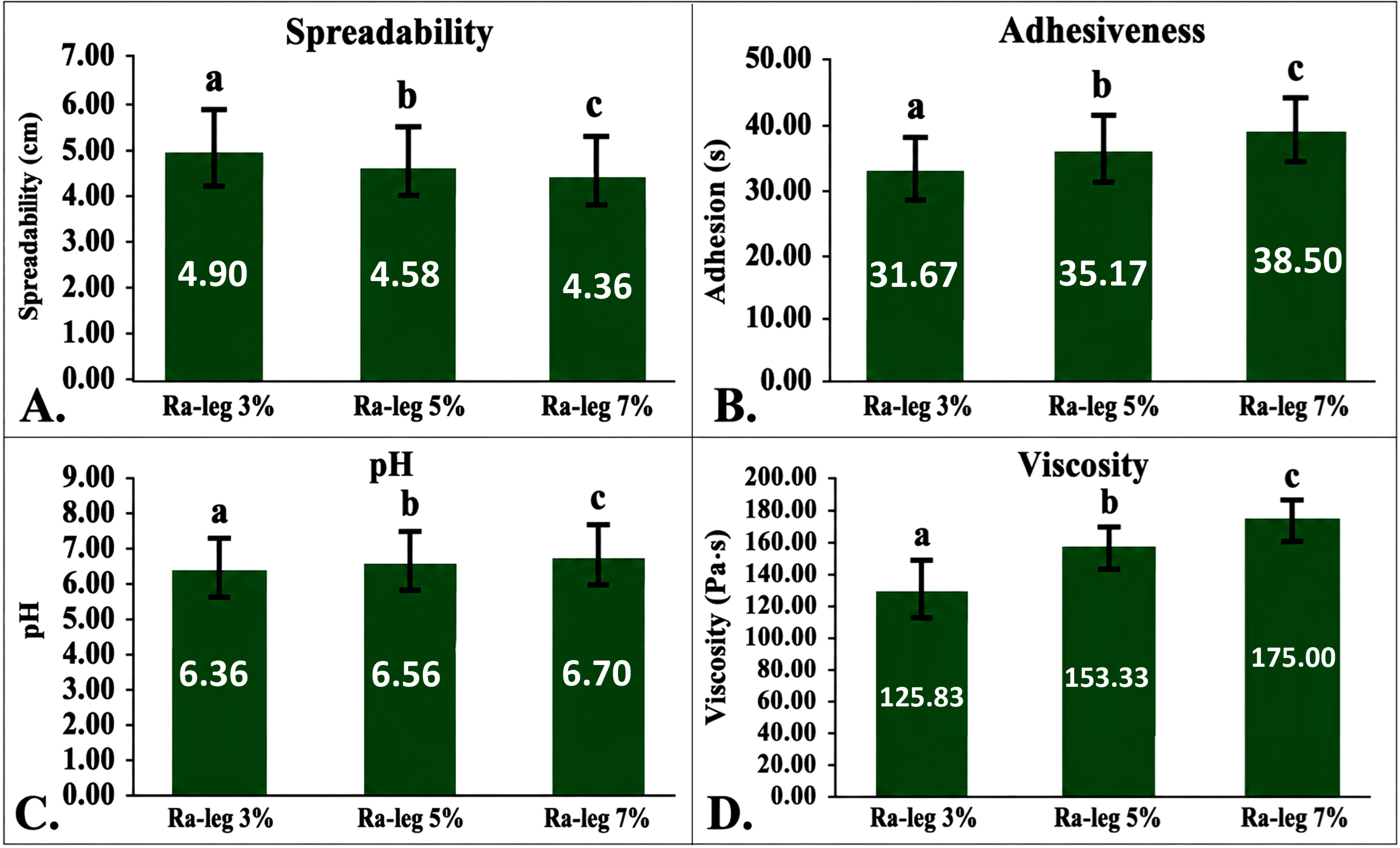
Physical characteristics of Ra-leg. (A) spreadability, (B) adhesiveness, (C) pH, and (D) viscosity of Ra-leg at concentrations of 3%, 5%, and 7%. Data are presented as mean ± SD. Different superscript letters indicate significant differences among groups (p < 0.05).
Figure 2 demonstrates the effect of R. apiculata Blume leaf extract concentration on the physical characteristics of Ra-leg formulations. A concentration-dependent pattern was observed across all evaluated parameters. Spreadability decreased significantly with increasing extract concentration, with the 3% formulation exhibiting the highest spreadability and the 7% formulation the lowest (p < 0.05). In contrast, adhesiveness, pH, and viscosity increased significantly as extract concentration increased (p < 0.05). Although pH increased slightly, all formulations remained within the physiologically acceptable range for topical application. The complete physical characterization data are presented in Table S1.
3.3. Flavonoid, Phenolic, and Tannin Levels
The standard curve used to measure the levels of QEF, GAEP, and TAET in Ra-leg is presented in Fig. (3).
Based on Figure 3, significant differences were observed in QEF, GAEP, and TAET among Ra-leg formulations at concentrations of 3%, 5%, and 7% (p < 0.05). The calibration curves for QEF, GAEP, and TAET showed good linearity across the tested concentration ranges, with coefficients of determination exceeding 0.98, indicating reliable analytical performance (Fig. 3A.1, B.1, and C.1). One-way ANOVA followed by Tukey’s post hoc test demonstrated a concentration-dependent increase in secondary metabolite contents (Fig. 3A.2, B.2, and C.2).
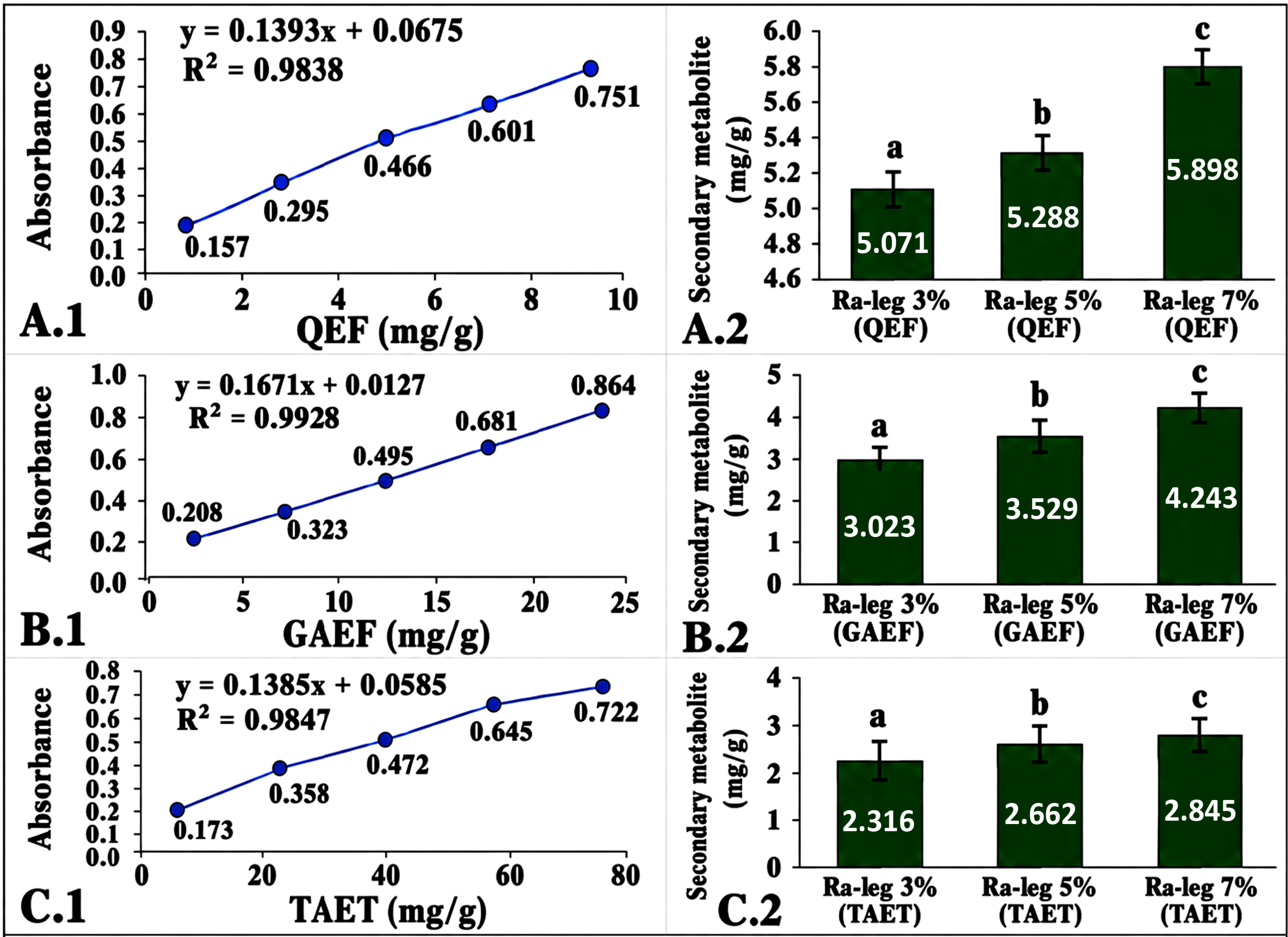
Calibration curves and secondary metabolite contents of Ra-leg. (A.1.) quercetin standard calibration curve for total flavonoid quantification (QEF); (B.1.) gallic acid standard calibration curve for total phenolic quantification (GAEP); (C.1.) tannic acid standard calibration curve for total tannin quantification (TAET). Absorbance was measured at 510 nm (QEF), 650 nm (GAEP), and 740 nm (TAET) using a UV–visible spectrophotometer. Bar graphs show the concentrations of (A.2.) QEF, (B.2.) GAEP, and (C.2.) TAET in Ra-leg formulations at concentrations of 3%, 5%, and 7%. Data are presented as mean ± SD (n = 5). Different superscript letters indicate significant differences among groups (p < 0.05), as determined by one-way ANOVA followed by Tukey’s post hoc test.
The contents of secondary metabolites increased with increasing extract concentration. Total flavonoid content (QEF) increased from 0.5082 ± 0.0015 mg QE/g gel in Ra-leg 3% to 0.5898 ± 0.0025 mg QE/g gel in Ra-leg 7%. The QEF values for all formulations are summarized in Table S2. Likewise, GAEP increased from 0.3175 ± 0.0075 mg GAE/g gel to 0.4395 ± 0.0067 mg GAE/g gel. GAEP data are provided in Table S3. TAET increased from 0.2316 ± 0.0014 mg TAE/g gel to 0.2845 ± 0.0025 mg TAE/g gel. TAE values are shown in Table S4.
3.4. Inhibition Zone Diameter of R. apiculata Blume Leaf Extract Gel against Bacteria
Photographs of the inhibition zone diameter of Ra-leg against bacteria are presented in Fig. (4).
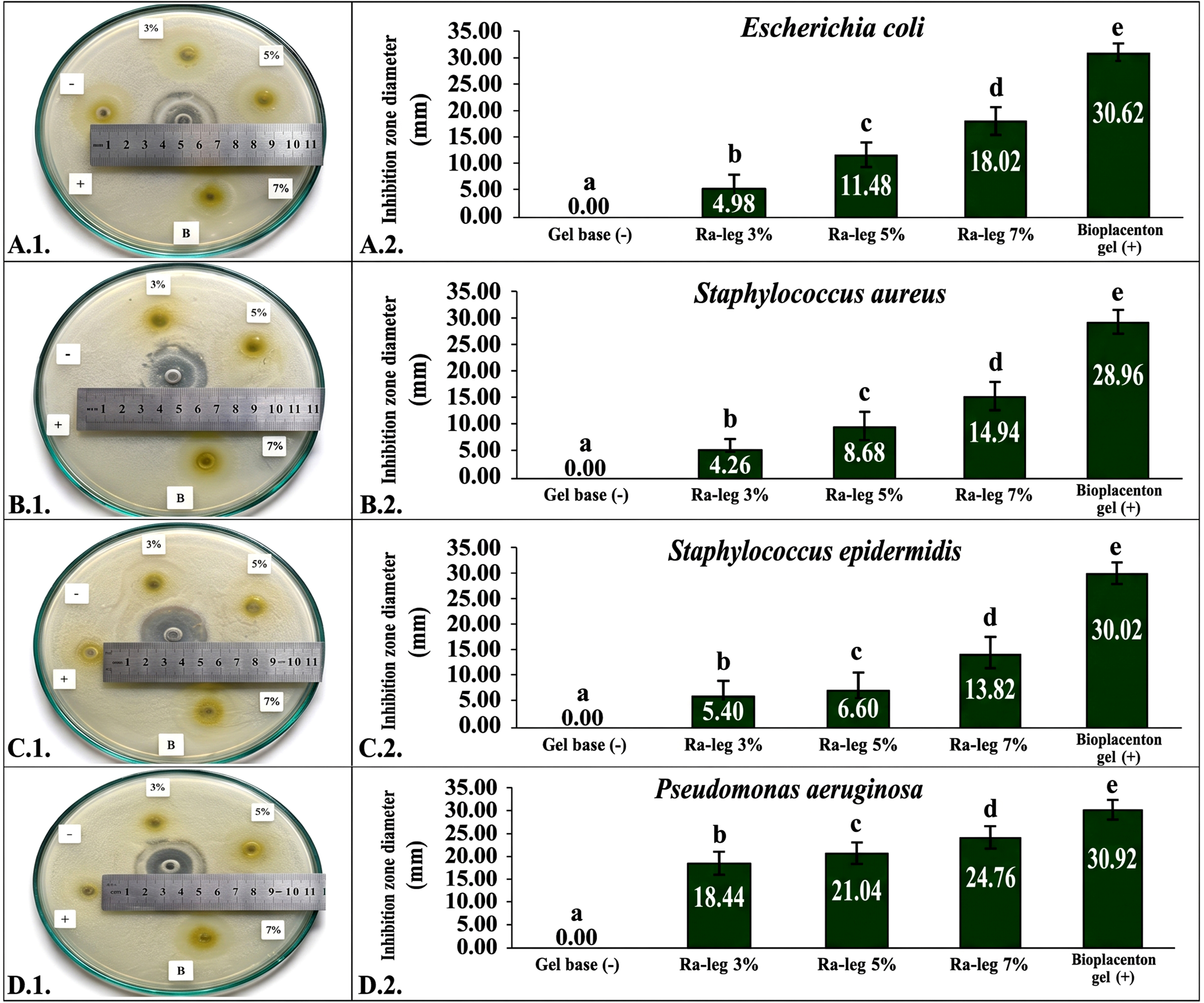
Antibacterial activity of Ra-leg. (A1) representative inhibition zones against Escherichia coli and (A2) corresponding quantitative data; (B1) representative inhibition zones against Staphylococcus aureus and (B2) corresponding quantitative data; (C1) representative inhibition zones against Staphylococcus epidermidis and (C2) corresponding quantitative data; (D1) representative inhibition zones against Pseudomonas aeruginosa and (D2) corresponding quantitative data. Data are presented as mean ± SD. Different lowercase letters (a-e) indicate statistically significant differences among groups (p < 0.05), as determined by one-way ANOVA followed by Tukey’s post hoc test.
The antibacterial activity of Ra-leg formulations against selected Gram-positive and Gram-negative bacteria is summarized in Fig. (4). The complete inhibition zone data are available in Table S5. The gel base (negative control) exhibited no inhibitory effect against any of the tested bacterial strains, confirming that the observed antibacterial activity was attributable to the incorporated extract. All Ra-leg formulations demonstrated significant, concentration-dependent antibacterial activity (p < 0.05). Increasing the extract concentration from 3% to 7% increased the inhibition zone diameters against all tested bacteria, with the 7% formulation producing the strongest antibacterial effect. Nevertheless, the inhibitory activity of Ra-leg at all concentrations remained significantly lower than that of the commercial Bioplacenton® gel used as a positive control (p < 0.05). Differences in bacterial susceptibility were also observed. P. aeruginosa exhibited the highest sensitivity to Ra-leg treatment, whereas S. epidermidis showed the lowest sensitivity at comparable concentrations. E. coli and S. aureus demonstrated intermediate and relatively similar susceptibility patterns.
4. DISCUSSION
The physicochemical properties of Ra-leg remained within normal and acceptable ranges, indicating that the gel formulation was suitable for topical application. These findings are consistent with previous studies on hydrogel-based drug delivery systems, which have highlighted the importance of appropriate physicochemical properties for skin compatibility and user acceptability [15]. In addition, organoleptic evaluation remains an important aspect of topical gel formulation development. The R. apiculata Blume leaf extract gel exhibited a clear appearance and semi-solid consistency, characteristics that are advantageous for biomedical applications [16].
The results of this study showed that the average spreadability diameter of Ra-leg ranged from 4.36 ± 0.08 to 4.90 ± 0.14 cm. According to established guidelines, an acceptable spreadability diameter for gel formulations is 4.0–7.0 cm. Other studies have also reported that spreadability values considered comfortable for semi-solid preparations range from 3–7 cm [17] and 5–7 cm [18]. Therefore, the spreadability of all Ra-leg formulations met the recommended criteria for topical gel preparations. In addition, the recommended adhesion time for gel formulations ranges from 10 to 180 s, with greater adhesion indicating improved drug retention on the skin [19]. By comparison, a Camellia sinensis sunscreen gel exhibited a considerably shorter adhesion time of approximately 0.68–1.46 s [20].
The results of this study indicate that the average pH of Ra-leg ranged from 6.36 ± 0.05 to 6.70 ± 0.05. This range falls within the acceptable pH limits for topical gel formulations (4.50–7.50). pH evaluation is essential to determine the potential of a gel to cause skin irritation upon application [9]. Normal human skin has a pH ranging from approximately 4.5 to 7.0 [15]. Previous studies on gels containing Ageratum conyzoides L. extract reported pH values between 5 and 6, with variations attributed to differences in secondary metabolite composition [17]. Other researchers have proposed narrower pH criteria, suggesting that gels with a pH close to the physiological skin pH are non-irritating [21]. Specifically, formulations with a pH range of approximately 4.5–6.5 have been reported to be compatible with the skin and well tolerated without inducing irritation [22]. Similarly, recent studies have shown that quercetin-loaded hydrogels exhibited a pH of 5.9 ± 0.03, which is considered suitable for wound healing. Such pH conditions (5.5–6.5) have been reported to be favorable for bacterial control, collagen synthesis, and the activation of enzymes involved in wound healing [23]. Therefore, careful control of gel pH during formulation is crucial, as extreme pH values may prolong gelation time and adversely affect formulation performance [24].
The acceptable average viscosity range for gel formulations is 120–180 Pa·s. Evaluation of Ra-leg viscosity is essential, as viscosity is an important rheological property influencing the performance and applicability of gel formulations. In addition to viscosity, other rheological parameters, such as viscoelasticity and elastic modulus, also contribute to gel characteristics [25]. The viscosity values obtained in this study differed from those reported for a Camellia sinensis leaf sunscreen gel, which exhibited a viscosity range of 123.33–203.33 Pa·s [20]. Notably, the increase in viscosity and adhesiveness observed at higher extract concentrations may improve gel retention on the skin surface, thereby supporting topical application of the formulation.
The QEF content of Ra-leg in this study ranged from 0.5082 ± 0.0015 to 0.5898 ± 0.0025 mg QE/g gel. This value is lower than the total flavonoid content reported for the ethanol extract of R. apiculata Blume leaves, which exhibited substantially higher concentrations in previous studies [26]. Likewise, the QEF content observed in this study was lower than the quercetin content (5.144%, equivalent to approximately 51.44 mg/g) identified in R. apiculata Blume leaves by HPLC analysis [27]. Comparable flavonoid levels have also been reported in Sargassum polycystum extracts, although the values varied depending on the extraction solvent and analytical method [28]. Variations in flavonoid content among different studies may be influenced by extraction conditions, particularly temperature, which is known to affect flavonoid stability and yield [29].
The GAEP content of Ra-leg ranged from 0.3175 ± 0.0075 to 0.4395 ± 0.0067 mg GAE/g gel. Previous studies reported higher total phenolic content in crude extracts of R. apiculata Blume. For example, the butanol fraction of the stem extract reached 181.84 mg GAE/g dry extract [30], whereas another study reported 53.24 ± 0.028 mg GAE/g extract [10]. The lower GAE values observed in Ra-leg are expected because phenolic compounds are dispersed within the gel matrix, resulting in lower concentrations per gram of formulation than in crude extracts. In addition, extraction solvent and plant part are known to influence phenolic yield [30]. Phenolic contents reported for Sargassum sp. extract (16.13 mg/g) were also higher than those observed in Ra-leg gel [28].
The TAET content of Ra-leg ranged from 0.2316 ± 0.0014 to 0.2845 ± 0.0025 mg TAE/g gel. Previous studies have reported that total tannin contents in the Rhizophoraceae family vary widely, ranging from 8.17 ± 1.36 mg/g to 813.35 ± 18.72 mg/g (p < 0.05) [31]. This variation reflects differences in plant species, plant parts, and extraction methods. Tannins are recognized as important bioactive phytochemicals, and tannin-rich extracts of R. apiculata Blume have been reported to exhibit significant antimicrobial potential [6]. In comparison, higher tannin contents have also been reported for Sargassum sp. extract (36.86 mg/g) [28].
The presence of flavonoids, phenolics, and tannins in the formulated Ra-leg is consistent with previous phytochemical investigations reporting similar secondary metabolite profiles in R. apiculata Blume leaves. These bioactive phytochemicals are known to contribute to antioxidant activity and are also associated with antibacterial mechanisms, including disruption of bacterial cell membranes, interaction with cellular proteins, and inhibition of microbial enzymatic systems. Therefore, the detected levels of QEF, GAEP, and TAET in the Ra-leg formulations may collectively contribute to the antibacterial activity observed against both Gram-positive and Gram-negative bacterial strains [1].
The inhibition zone diameters of Ra-leg against E. coli differed significantly among treatment groups (p < 0.001). Compared with the positive control (Bioplacenton® gel), all Ra-leg formulations produced significantly smaller inhibition zones against E. coli (p < 0.001). Similarly, previous studies have reported that gallic acid nanocrystal hydrogel exhibited a larger inhibition zone against E. coli (21.52 ± 0.06 mm) than those observed for Ra-leg at all tested concentrations [32].
The inhibition zone diameters of Ra-leg against S. aureus differed significantly among treatment groups (p < 0.001). Compared with the positive control (Bioplacenton® gel), all Ra-leg formulations produced significantly smaller inhibition zones against S. aureus (p < 0.001). Similarly, previous studies reported that gallic acid nanocrystal hydrogel exhibited a larger inhibition zone against S. aureus (39.75 ± 0.61 mm) than those observed for Ra-leg formulations [32]. Previous studies have also demonstrated the antibacterial activity of plant-based topical hydrogel formulations against S. aureus. These differences indicate that antibacterial activity may vary according to the plant extract used and the physicochemical characteristics of the hydrogel formulation [33].
The inhibition zone diameters of Ra-leg against S. epidermidis differed significantly among treatment groups (p < 0.001). Compared with the positive control (Bioplacenton® gel), all Ra-leg formulations produced significantly smaller inhibition zones against S. epidermidis (p < 0.001). Previous studies reported that a gel containing palm oil (Elaeis guineensis Jacq.) exhibited inhibition zone diameters ranging from 9.61 to 11.60 mm against S. epidermidis. Compared with these values, the Ra-leg formulations at 3% and 5% produced smaller inhibition zones, whereas the 7% formulation exhibited a larger inhibition zone, indicating higher antibacterial activity against S. epidermidis [34].
The inhibition zone diameters of Ra-leg against P. aeruginosa differed significantly among treatment groups (p < 0.001). Compared with the positive control (Bioplacenton® gel), all Ra-leg formulations produced significantly smaller inhibition zones against P. aeruginosa (p < 0.001). In the present study, the 7% Ra-leg formulation produced an inhibition zone of 24.76 ± 0.21 mm against P. aeruginosa, which was greater than the inhibition zone of up to 18.62 mm reported for crude bark extracts of R. apiculata Blume [35]. This difference reflects the use of different plant materials (bark versus leaves) and different dosage forms (crude extract versus gel formulation).
The concentration-dependent increase in antibacterial activity observed in Ra-leg was accompanied by increased levels of flavonoids, phenolics, and tannins. These findings support the potential of Ra-leg as a bioactive gel with measurable antibacterial properties for further development as a topical therapeutic formulation.
Although the antibacterial activity of Ra-leg was most pronounced against P. aeruginosa, moderate inhibitory effects were also observed against E. coli and S. aureus. In contrast, S. epidermidis exhibited the lowest susceptibility among the tested bacterial strains. These findings indicate differences in the antibacterial activity of Ra-leg against the tested bacteria and support its potential for further development as a topical antibacterial formulation.
Importantly, R. apiculata Blume leaf utilization does not threaten species sustainability. Selective leaf harvesting preserves tree structure and supports natural regeneration. R. apiculata Blume is widely distributed along the Indonesian coastline. The species also exhibits strong natural regenerative capacity. These characteristics support sustainable scientific and industrial utilization under responsible harvesting practices.
5. STUDY LIMITATION
This study has several limitations. Ra-leg was not compared with the crude R. apiculata Blume leaf extract or individual secondary metabolites. Furthermore, only in vitro antibacterial activity was evaluated. Therefore, further in vivo studies and clinical trials are required to confirm the therapeutic efficacy and safety of Ra-leg formulations.
CONCLUSION
This study reports the first development of a standardized topical gel from R. apiculata Blume leaf extract. R. apiculata Blume leaf extract gel (Ra-leg) formulations at concentrations of 3%, 5%, and 7% exhibited acceptable physical and chemical characteristics for topical application. All formulations were semi-solid, homogeneous, transparent yellowish-green in appearance, and possessed the characteristic odor of the extract. The gels demonstrated spreadability ranging from 4.3 to 4.9 cm, adhesiveness between approximately 31 and 38.5 s, pH values within the physiologically acceptable range for skin application (6.3–6.7), and viscosity values increasing from 125 to 175 Pa·s.
The contents of QEF, GAEP, and TAET increased significantly with extract concentration, following the order Ra-leg 3% < 5% < 7% (p < 0.05). This finding indicates a positive correlation between extract concentration and secondary metabolite content. All Ra-leg formulations exhibited antibacterial activity against E. coli, S. aureus, S. epidermidis, and P. aeruginosa, although the inhibition zones were smaller than those produced by the positive control (Bioplacenton® gel). The strongest antibacterial effect was consistently observed in the 7% Ra-leg formulation across all tested bacterial strains. Overall, these findings suggest that Ra-leg, particularly at higher concentrations, possesses favorable physicochemical properties and concentration-dependent antibacterial activity, supporting its potential development as a topical antibacterial formulation. These findings provide a foundational framework for the advancement of Ra-leg as a standardized, plant-based therapeutic for managing skin infections.
AUTHORS’ CONTRIBUTIONS
The authors confirm their contributions to the paper as follows: E.X., D.T., and H.J.E.: Conceived and designed the study; E.X., D.T., H.J.E., J.J.T., and L.G.: Participated in data collection, data analysis, and interpretation of the results; E.X., J.J.T., L.G., and H.J.E.: Reviewed and processed the images. E.X., D.T., H.J.E., A.V.O., and S.K.: Contributed to manuscript preparation and writing. All authors reviewed, revised, and approved the final version of the manuscript.
LIST OF ABBREVIATIONS
| Ra-leg | = Rhizophora apiculata Blume leaf extract gel |
| QEF | = Quercetin Equivalent Flavonoid |
| GAEP | = Gallic Acid Equivalent Phenolic |
| TAET | = Tannic Acid Equivalent Tannin |
| QE | = Quercetin Equivalent |
| GAE | = Gallic Acid Equivalent |
| TAE | = Tannic Acid Equivalent |
| UV-Vis | = Ultraviolet-Visible spectrophotometry |
| HPMC | = Hydroxypropyl Methylcellulose |
| PG | = Propylene glycol |
| TEA | = Triethanolamine |
| q.s. | = quantum satis (sufficient quantity) |
| Pa·s | = Pascal second |
| cm | = centimeter |
| s | = second |
| mm | = millimeter |
| mg | = milligram |
| g | = gram |
| mL | = milliliter |
| °C | = degrees Celsius |
| SD | = Standard Deviation |
| ANOVA | = Analysis of Variance |
| HSD | = Honestly Significant Difference |
| IUCN | = International Union for Conservation of Nature |
| CITES | = Convention on International Trade in Endangered Species |
| LPPT | = Laboratorium Penelitian dan Pengujian Terpadu |
| HPLC | = High-Performance Liquid Chromatography |
ETHICS APPROVAL AND CONSENT TO PARTICIPATE
This study was approved by the Research Ethics Commission, Faculty of Medicine, Universitas Trisakti, Jakarta, Indonesia (RSH/NK/INT/202504/0001).
RESEARCH INVOLVING PLANTS
The plant material used in this study was R. apiculata Blume. The species is not listed as endangered under the IUCN Red List (category: Least Concern) [36]. It is widely distributed in Southeast Asian mangrove ecosystems [37]. The collection and utilization of the plant followed relevant institutional guidelines. The procedures also complied with international principles for the ethical use of plant resources, including the IUCN Policy Statement on Research Involving Species at Risk of Extinction.
AVAILABILITY OF DATA AND MATERIALS
The data and materials utilized in this study are available within the published article.
FUNDING
The publication of this research manuscript was funded by the Faculty of Medicine, Universitas Trisakti, Jakarta, Indonesia, with the number 0936/PUF/FK/ 2024-2025
ACKNOWLEDGEMENTS
Gratitude is expressed to the Head and Staff of the Integrated Research and Testing Laboratory (Laboratorium Penelitian dan Pengujian Terpadu = LPPT) of Universitas Gadjah Mada, so that this research to be carried out. Special thanks are also extended to Marlita Sabta Utari and Mellysa Henggar Ratnasari for their valuable contributions to data collection and image processing.